Interventional Cardiology Procedures

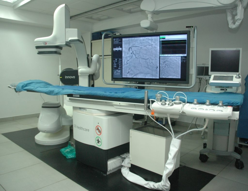
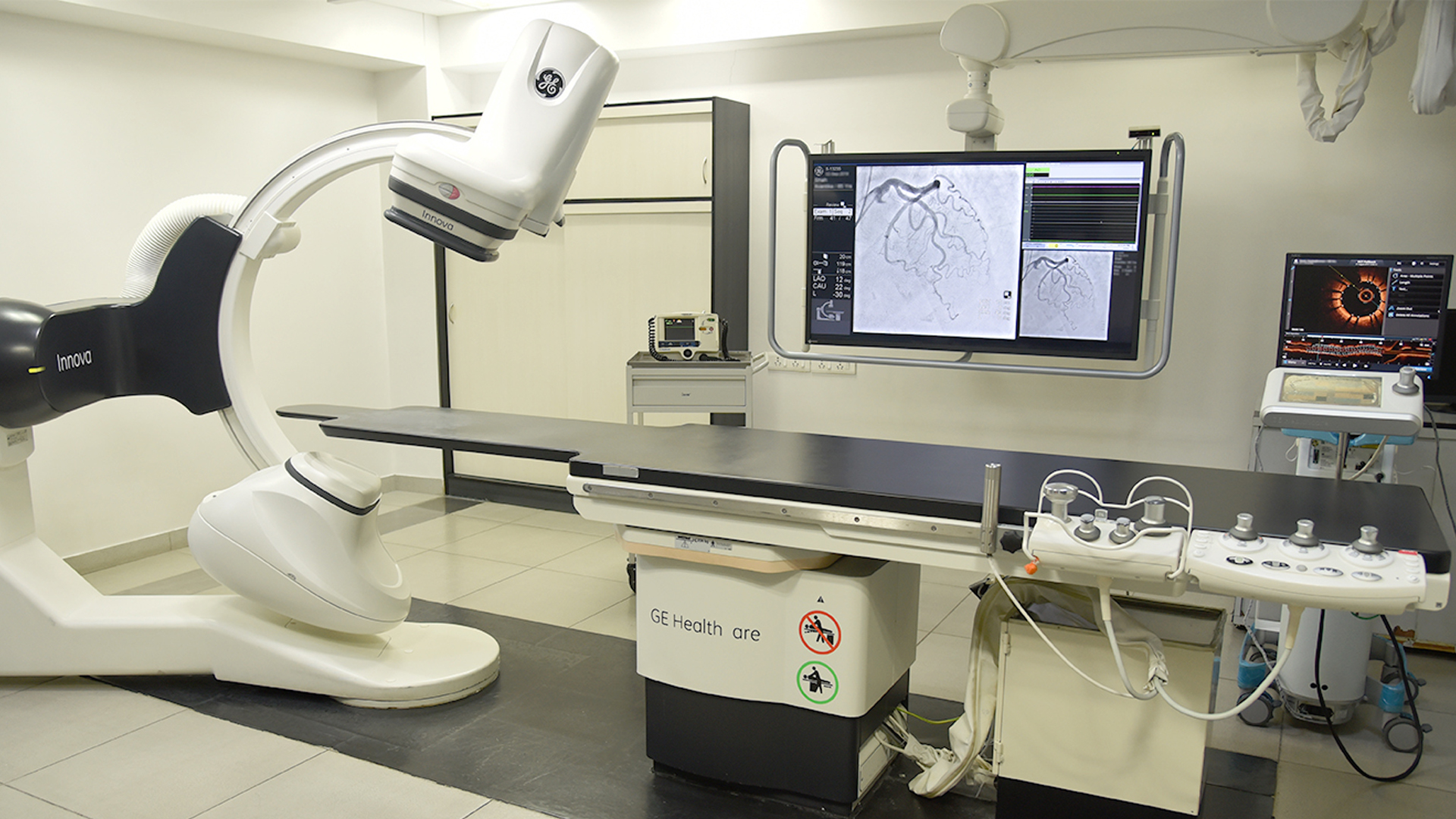
Technology Used In Cardiac Cath Lab
Sometimes the fatty substance that builds up in arteries (plaque) contains calcium that makes the blockage hard. If a plaque is severely calcified, a standard angioplasty balloon may not be able to cross the blockage and push it to the sides of the artery.Rotational atherectomy is a procedure that can be performed to drill through tough blockages. A tiny rotating cutting device is used to open a narrowed artery and improve blood flow. The pieces of plaque dislodged by the rotational atherectomy device are small enough to be absorbed by the blood stream.
FFR is a test used to measure how much blood flow is being restricted by a blockage in an artery. A special, pressure-sensing guidewire is fed through a catheter to the site of the blockage in the artery.
When water flows through a garden hose the flow is driven by the pressure in the tap. If there hose has no obstruction, the pressure at the end of the hose is the same as the pressure at the tap. In a healthy heart artery, the pressure at the end of the artery is the same as at the beginning of the artery (where it comes off of the aorta). But when a blockage reduces flow through the heart artery, or a garden hose, the pressure at the end is reduced proportionate to the restriction. The greater the restriction, the lower the pressure downstream because the flow is reduced. When the FFR wire is placed across the blockage, it measures the pressure in front of and beyond the blockage. If the blood pressure in the artery or beyond the blockage is found to be significantly reduced, then that blockage may be a good candidate for angioplasty and stenting to clear the blockage and prop the artery open.
All ultrasound tests, including IVUS, use sound waves to create images. IVUS is used to gather images of the inside of arteries to find out if a blockage is present, and if so, how serious the blockage is.
During an IVUS test, a catheter with an ultrasound probe at the end is threaded over a guidewire in the artery to the area to be tested. The ultrasound catheter sends out sound waves and receives echoes from the sound waves as they bounce back from the body’s tissues. These echoes are translated by a computer into images of the artery. IVUS is a test that may be performed during an angiogram (also known as cardiac catheterization). Your doctor may use IVUS if the blockages seen with the angiogram appear to be borderline-severe, or if the doctor needs more information about the plaque anatomy.
IVUS is useful because it allows the interventional cardiologist to measure the amount of plaque inside vessels as well as how much much space is available in the artery for blood to flow through. If it is determined that you need angioplasty to treat the blocked artery, IVUS can help with accurate positioning of the balloon and stent.
OCT is an imaging tool used to take high-resolution pictures of blood vessel walls. OCT provides interventional cardiologists with detailed images of plaque (cholesterol and other materials that have accumulated in the walls of the artery and can rupture, causing a blood clot to form at the site and block off critical blood flow). Like IVUS, this detailed information about plaque build-up in arteries can help interventional cardiologists determine where best to place stent
Shockwave intravascular lithotripsy (IVL) is an innovative medical technique adapted from the established treatment for kidney and ureteral stones. It involves the use of a percutaneous device to generate acoustic pressure waves, which are directed towards calcified lesions in blood vessels. These waves effectively break down both superficial and deep calcium deposits within the vessel walls. This process assists in preparing the artery for the successful deployment of a vascular stent, ultimately restoring proper blood flow and improving patient
Non Invasive Cardiology Tests


